.
.
KEY TO SPECIES OF APHODIINAE OF SOUTHERN SOUTH AMERICA

(From: Smith, A. B. T. and P. E. Skelley. 2007. A review of the Aphodiinae (Coleoptera: Scarabaeidae)
of southern South America. Zootaxa 1458: 1-80).

.
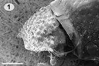
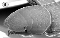
1. Head strongly convex, clypeal surface rugosely punctate and distinctly setose
(Fig. 1) ................................. Argeremazus neuquen Stebnicka and Dellacasa

- Head flat to weakly convex; clypeal surface variable, smooth to tuberculate, finely to coarsely punctate, sometimes weakly setose, but never convex and distinctly setose (Figs. 2–5) ......................................................................................... 2

clypeus clypeus


Fig. 1. Argeremazus neuquen, head; Fig. 2. Aegialia argentina head to elytral base;
Fig. 3. Ataenius platensis head to elytral base; Fig. 4. Aphodius granarius head to elytral base;
Fig. 5. Pleurophorus caesus head to elytral base. Scale line = 0.2 mm.
.
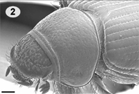
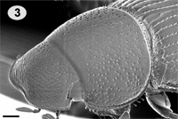
2. Labrum and mandibles not visible in antero-dorsal view, hidden beneath expanded clypeus (Figs. 3–5) ......................................................................... 3

- Labrum and mandibles visible in antero-dorsal view (Fig. 2) [Aegialiini] ................................................................................................................... 7
.
clypeus clypeus  
     
clypeus clypeus  
     
Fig. 2. Aegialia argentina head to elytral base; Fig. 3. Ataenius platensis head to elytral base; Fig. 4. Aphodius granarius head to elytral base;
Fig. 5. Pleurophorus caesus head to elytral base. Scale line = 0.2 mm.
.
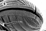
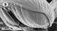
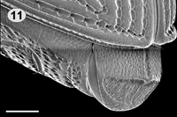
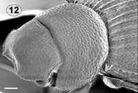
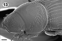
3. Elytral intervals margined at base (most visible toward the sides) (Figs. 3, 5, 12, 13); pygidium with basal longitudinal groove (Figs. 6, 11), usually eroded in apical half (Fig. 10); elytra with 
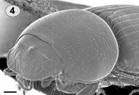
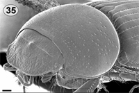
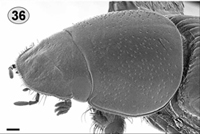
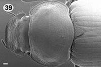
internal swelling along sutural margin that fits into the pygidial groove (Fig. 6) ................................................................................. 4

- Elytral intervals not margined at base (Figs. 2, 4), smoothly rounded (Figs. 35, 36, 39); pygidium entirely smooth (Fig. 9), unmodified, never eroded in apical half; elytral apex at suture not enlarged, sharply edged (Fig. 9) ................................ 5

clypeus clypeus
     
clypeus clypeus elytra
     
elytra clypeus
     
clypeus clypeus clypeus
 
Fig. 1. Argeremazus neuquen, head; Fig. 2. Aegialia argentina head to elytral base;
Fig. 3. Ataenius platensis head to elytral base; Fig. 4. Aphodius granarius head to elytral base;
Fig. 5.
Pleurophorus caesus head to elytral base; Fig. 6. Ataenius picinus apex of abdomen and elytra; Fig. 9. Aidophus parcus apex of abdomen and elytra; Fig. 11. Ataenius platensis pygidium; Fig. 12. Bruchaphodius shannoni head to elytral base; Fig. 13. Parataenius simulator head to elytral base; Fig. 35. Aphodius granarius head to elytral base; Fig. 36. Aphodius pseudolividus head to elytral base. Fig. 39. Podotenus (Pseudopodotenus) fulviventris head to elytral base;
Scale line = 0.2 mm.
.
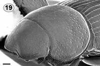
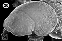
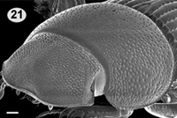
4. Clypeus smooth, with transverse wrinkles or transverse ridges (Figs. 3, 12–15, 19–21); pronotum without 
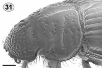
grooves or rows of punctures; metafemur never greatly swollen, not larger than profemur [Eupariini] ................................................... 8

- Clypeus granulate or tuberculate, never with transverse ridges (Figs. 5, 28, 29, 31–33); pronotum usually with rows of punctures; metafemur usually enlarged (not in Pleurophorus), larger than profemur (Fig. 30) [Psammodiini] ........................ 18

clypeus clypeus
     
clypeus clypeus clypeus


Fig. 3. Ataenius platensis head to elytral base; Fig. 5. Pleurophorus caesus head to elytral base; Fig. 12. Bruchaphodius shannoni head to elytral base; Fig. 13. Parataenius simulator head to elytral base; Fig. 14. Ataenius gracilis head; Fig. 15. Ataenius chilensis head; Fig. 19. Ataenius opatroides head to elytral base; Fig. 20. Ataenius picinus head to elytral base; Fig. 21. Ataenius strigicaudus head to eltyral base; Fig. 28. Odontopsammodius cruentus head to elytral base;
Fig. 29.
Tesarius caelatus head to elytral base; Fig. 31. Leiopsammodius indefensus head to elytral base; Fig. 32. Leiopsammodius placidus (holotype) head to elytral base; Fig. 33. Platytomus micros head to elytral base. Scale line = 0.2 mm.
.

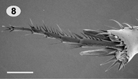
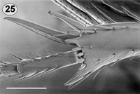
5. Metatibia with apical spurs not separated by metatarsus (Fig. 7) ..................... 6

- Metatibia with apical spurs separated by metatarsus (Fig. 8) [Aphodiini] .......... 23

   
Fig.7. Aidophus infuscatopennis metatibial apex, ventral view;
Fig. 8. Aphodius rubeolus metatibial apex, ventral view. Scale line = 0.2 mm.
.

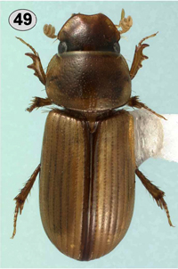
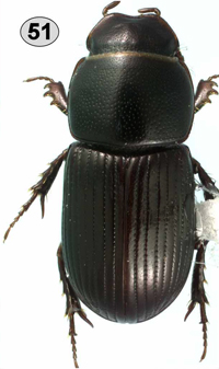
6. Body robust, moderately elongate, dark brown without markings, resembling a small A. granarius (Fig. 51) [Proctophanini]
............................................................ Australaphodius frenchi (Blackburn)

- Body not robust, distinctly elongate and parallel-sided, yellow brown with darker markings (Figs. 49–50) [Didactyliini]
............................................ Aidophus spp. [see Erroneous Records section]

     
Fig. 49. Aidophus flaveolus habitus; Fig. 50. Aidophus infuscatopennis habitus;
Fig. 51. Australaphodius frenchi habitus. Scale line = 0.2 mm
.
AEGIALIINI

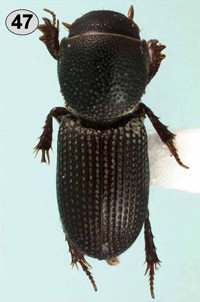
7. Body elongate, black (Fig. 47); metatibia with apical spurs not separated by tarsus ........................................ Amerisaprus valdivia Stebnicka and Skelley

- Body robust, reddish-brown (Fig. 46); metatibia with apical spurs separated by tarsus ............................... Aegialia argentina Martínez, Pereira, and Vulcano

   
Fig. 47. Amerisaprus valdivia habitus; Fig. 48. Argeremazus neuquen habitus. Scale line = 0.2 mm
.
EUPARIINI
8. Clypeal apex distinctly dentate (Fig. 12); body robust, dark red-brown ............ 9

- Clypeal apex evenly rounded at sides, or weakly angulate, never dentate (Figs. 13–15); body usually elongate and black or red ............................................... 10
   
   
Fig. 12. Bruchaphodius shannoni head to elytral base.
Fig. 13.
Parataenius simulator head to elytral base;
Fig. 14. Ataenius gracilis head; Fig. 15. Ataenius chilensis head.
Scale line = 0.2 mm
.

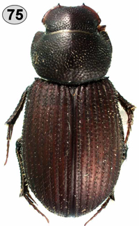
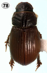
9. Elytral intervals flattened with 2 rows of distinct setae (Fig. 75) ............................................................. Bruchaphodius ovalipennis (Harold)
- Elytral intervals with distinct median ridge, lacking setae (Figs. 12, 76) ................................................................. Bruchaphodius shannoni (Bruch)

     
Fig. 12. Bruchaphodius shannoni head to elytral base. Fig. 75. Bruchaphodius ovalipennis habitus; Fig. 76. Bruchaphodius shannoni habitus. Scale line = 0.2 mm
.
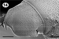
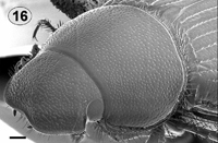
10. Clypeal surface with distinct, strongly developed, transverse ridges (Figs. 13, 16); mesotibia strongly 
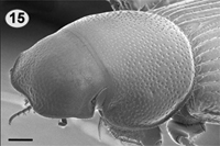
expanded at apex; metafemur swollen, nearly as large as profemur .................................................................................................... 11

- Clypeal surface variable, punctate, granulate, or with transverse wrinkles, never with distinct ridges (Figs. 14–15); mesotibia not strongly expanded at apex; metafemur not or weakly swollen, smaller than profemur ................................ 12

   
Fig. 13. Parataenius simulator head to elytral base;
Fig. 14. Ataenius gracilis head; Fig. 15. Ataenius chilensis head; Fig. 16. Parataenius derbesis head to elytral base. Scale line = 0.2 mm
.

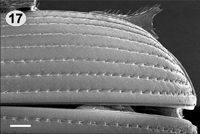
11. Coarse pronotal punctures restricted to postero-lateral third and base, very much larger than other punctures, rapidly decreasing in size anteriorly at middle (Fig. 13); punctures of elytral intervals fine and widely spaced (Fig. 17) ..................................................................... Parataenius simulator (Harold)

- Coarse pronotal punctures evenly distributed across surface, not as notably different from smaller punctures (Fig. 16); punctures of elytral intervals dense and restricted to central part of interval (Fig. 18)
.................... Parataenius derbesis (Solier) [see Erroneous Records section]

   
Fig. 13. Parataenius simulator head to elytral base;
Fig. 16.
Parataenius derbesis head to elytral base;
Fig. 17. Parataenius simulator elytral base; Fig. 18. Parataenius derbesis elytral base. Scale line = 0.2 mm
.
12. Body distinctly setose and densely, coarsely punctate ...................................................................... Oxyataenius morosus (Harold)

- Body neither distinctly hairy nor densely, coarsely punctate ............................ 13
.

13. Clypeal surface coarsely and densely punctate, punctures elongate (Fig. 14)
....................................................................... Ataenius gracilis (Melsheimer)

- Clypeal surface variable, but punctures never elongate (Figs. 15, 19–21) ......... 14


Fig. 14. Ataenius gracilis head; Fig. 15. Ataenius chilensis head; Fig. 19. Ataenius opatroides head to elytral base; Fig. 20. Ataenius picinus head to elytral base; Fig. 21. Ataenius strigicaudus head to eltyral base. Scale line = 0.2 mm
.

14. Clypeal surface weakly granulate on apical half (Fig. 15) ............................................................................. Ataenius chilensis (Solier)

- Clypeal surface punctate, smooth or weakly wrinkled (Figs. 19–21) .................. 15

   
Fig. 15. Ataenius chilensis head; Fig. 19. Ataenius opatroides head to elytral base; Fig. 20. Ataenius picinus head to elytral base; Fig. 21. Ataenius strigicaudus head to eltyral base.
Scale line = 0.2 mm
.

15. Head smooth, apparently without punctures (Fig. 19); pronotum apparently lacking fine punctures, coarse punctures present laterally (Fig. 19); pronotum and elytra dull .................................................... Ataenius opatroides (Blanchard)

- Head with distinct punctation and often with transverse wrinkles (Figs. 3, 20–21); pronotum distinctly punctate throughout with dense fine punctures, coarse punctures also present laterally (Figs. 3, 20–21); pronotum and elytra shiny ...... 16

   
Fig. 3. Ataenius platensis head to elytral base; Fig. 19. Ataenius opatroides head to elytral base; Fig. 20. Ataenius picinus head to elytral base; Fig. 21. Ataenius strigicaudus head to eltyral base.
Scale line = 0.2 mm
.

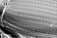
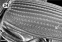
16. Elytral interval 9 (penultimate lateral interval) weakly punctate (Fig. 22), but not different from those of disc; pronotum with marginal setae near posterior angle spatulate (Fig. 3), flattened and widest near apex; meso and metatibial accessory spine near base of apical spurs short, at most as long as 4–6 apical spinules
...................................................................... Ataenius platensis (Blanchard)

- Elytral interval 9 (penultimate lateral interval) with puncture pattern differing from those of disc (Figs. 23–24); pronotum with marginal setae near posterior angle not notably spatulate, possibly flattened, but not widest near apex; meso and metatibial accessory spine near base of apical spurs longer than 4 apical spinules (Fig. 25) .. 17


Fig. 3. Ataenius platensis head to elytral base; Fig. 22. Ataenius platensis lateral elytron;
Fig. 23.
Ataenius picinus lateral elytron; Fig. 24. Ataenius strigicaudus lateral elytron;
Fig. 25.
Ataenius picinus apex of metatibia. Scale line = 0.2 mm.
.

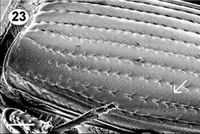
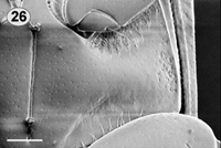
17. Elytral interval 9 with fine, dense punctures covering entire surface (Fig. 23); metasternum lacking coarse punctures medially (Fig. 26) ................................................................................. Ataenius picinus Harold

- Elytral interval 9 with 2 rows of coarse punctures (Fig. 24); metasternum with group of coarse punctures near base of mesofemur (Fig. 27) ........................................................................... Ataenius strigicaudus Bates

   
Fig. 23. Ataenius picinus lateral elytron; Fig. 24. Ataenius strigicaudus lateral elytron; Fig. 26. Ataenius picinus metasternum; Fig. 27. Ataenius strigicaudus
metasternum. Scale line = 0.2 mm.
.
PSAMMODIINI

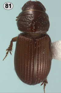
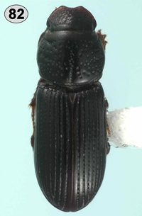
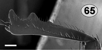
18. Body elongate, cylindrical, parallel-sided for majority of length (Fig. 82); metafemur parallel-sided, not swollen ................. Pleurophorus caesus (Panzer)

- Body more robust, not elongate (Figs. 78–81, 83); metafemur swollen, at most weakly parallel-sided .................................................................................... 19


Fig. 78. Leiopsammodius indefensus habitus; Fig. 79. Leiopsammodius placidus habitus (holotype); Fig. 80. Odontopsammodius cruentus habitus;
Fig. 81. Platytomus micros habitus; Fig. 82. Pleurophorus caesus habitus;
Fig. 83.
Tesarius caelatus habitus.

.

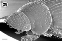
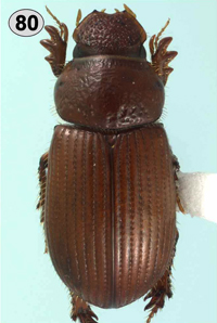
19. Clypeal apex distinctly bidentate (Figs. 28, 80) ......................................................... Odontopsammodius cruentus (Harold)

- Clypeal apex at most angulate, usually evenly sinuate (Figs. 29, 31–32) .......... 20

   
Fig. 28. Odontopsammodius cruentus head to elytral base; Fig. 29. Tesarius caelatus head to elytral base; Fig. 31. Leiopsammodius indefensus head to elytral base; Fig. 32. Leiopsammodius placidus (holotype) head to elytral base. Scale line = 0.2 mm.
Fig. 80. Odontopsammodius cruentus habitus.
.

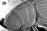
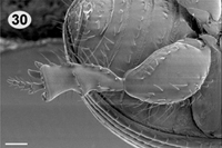
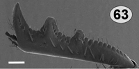
20. Metatibia with complete transverse ridge near middle (Fig. 30); eyes reduced (Fig. 29); flightless .............................................. Tesarius caelatus (LeConte)

- Metatibia lacking complete transverse ridge, possibly with transverse row of teeth; eyes normally developed (Figs. 31–32); flightless or not .................................. 21

   
Fig. 29. Tesarius caelatus head to elytral base;
Fig. 30. Tesarius caelatus metafemur and tibia;
Fig. 31.
Leiopsammodius indefensus head to elytral base;
Fig. 32. Leiopsammodius placidus (holotype) head
to elytral base. Scale line = 0.2 mm.
.

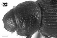
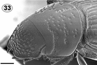
21. Elytra lacking setae on lateral margin; base of head roughly punctate, lacking grooves (Figs. 32–33); pronotum grooves weak .............................................. 22

- Elytra with distinct, long setae on lateral margin (Fig. 78); base of head with angled grooves (Figs. 31, 78); pronotum with one longitudinal groove on midline and 2 transverse grooves on each side (Figs. 31, 78) ......................................................... Leiopsammodius indefensus (Schmidt)

   
Fig. 31. Leiopsammodius indefensus head to elytral base;
Fig. 32.
Leiopsammodius placidus (holotype) head to elytral base; Fig. 33. Platytomus micros head to elytral base. Scale line = 0.2 mm. Fig. 78. Leiopsammodius indefensus habitus.
.

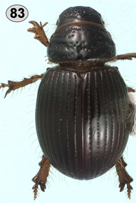
22. Body more elongate, elytra parallel-sided (Fig. 81); clypeus rounded on each side of central emargination; pronotal lateral margin lacking fringe of setae (Fig. 33) ............................................................................ Platytomus micros (Bates)

- Body more globose, elytra not parallel-sided (Fig. 79); clypeus angulate on each side of central emargination; pronotal lateral margin with distinct fringe of setae (Fig. 32) ................................................ Leiopsammodius placidus (Schmidt)

   
Fig. 32. Leiopsammodius placidus (holotype) head to elytral base; Fig. 33. Platytomus micros head to elytral base. Scale line = 0.2 mm. Fig. 79. Leiopsammodius placidus habitus (holotype);
Fig. 81.
Platytomus micros habitus.
.
APHODIINI

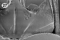
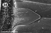
23. Scutellum narrowed at base, pentagonal (Fig. 34); head with tubercles on frontal suture (Figs. 35– 36) ........................................................................ 24

- Scutellum broadly triangular, not narrowed at base; head lacking tubercles on frontal suture (Figs. 39, 58– 62, 66–68) ........................................................ 25

     
Fig. 34. Aphodius granarius scutellum; Fig. 35. Aphodius granarius head to elytral base;
Fig. 36. Aphodius pseudolividus head to elytral base; Fig. 39. Podotenus (Pseudopodotenus) fulviventris head to elytral base. Fig. 58. Orodaliscoides reflexus male habitus;
Fig. 59. Orodaliscoides reflexus female habitus; Fig. 60. Orodaliscoides rugosiceps habitus;
Fig. 61. Acanthaphodius bruchi habitus; Fig. 62. Symphodon anomalus habitus;
Fig. 66. Podotenus (Paranimbus) sp. male habitus; Fig. 67. Podotenus fulviventris male habitus; Fig. 68. Podotenus fulviventris female habitus. Scale line = 0.2 mm.
.

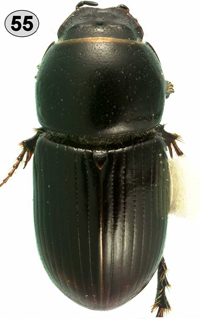
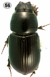
24. Pronotum with distinct, complete basal margin (Figs. 35, 55–56); body black
......................................... Aphodius (Calamosternus) granarius (Linnaeus)

- Pronotum lacking basal margin (Fig. 36); body bicolored (Fig. 57) .............................................. Aphodius (Labarrus) pseudolividus Balthasar

     
Fig. 35. Aphodius granarius head to elytral base; Fig. 36. Aphodius pseudolividus head to elytral base. Scale line = 0.2 mm. Fig. 55. Aphodius granarius male habitus; Fig. 56. Aphodius granarius female habitus; Fig. 57. Aphodius pseudolividus habitus.
.

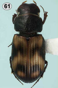
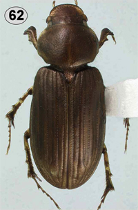
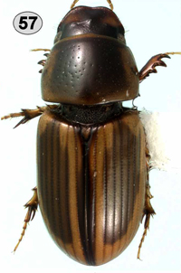
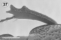
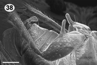
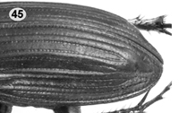
25. Small, less than 5 mm; protibia with apical most tooth projecting forward (Fig. 37); elytra with distinct color pattern (Fig. 61); male metafemur with distinct medial peg (Fig. 38) ................................................ Acanthaphodius bruchi Schmidt

- Larger, greater than 6 mm; protibia with apical most tooth projecting laterally (Figs. 40–42, 63–65); elytra unicolorous, or with paler apex only; male metafemur lacking peg ............................................................................................... 26


Fig. 37. Acanthaphodius bruchi protibia; Fig. 38. Acanthaphodius bruchi metafemur male;
Fig. 40.
Orodaliscoides reflexus protibia; Fig. 41. Orodaliscoides rugosiceps protibia;
Fig. 42. Symphodon anomalus protibia; Fig. 61. Acanthaphodius bruchi habitus;
Fig. 63.
Podotenus fulviventris female protibia; Fig. 64. Podotenus fulviventris male protibia;
Fig. 65.
Podotenus (Paranimbus) sp. male protibia. Scale line = 0.2 mm.
.

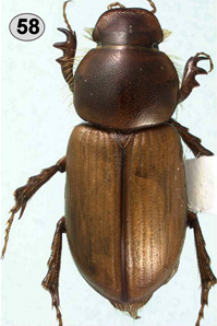
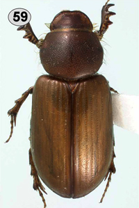
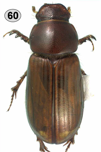
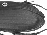
26. Body stout, head convex, clypeal margin strongly reflexed (Figs. 58–60); elytra evenly rounded apically, without apical umbone (Fig. 3) .................................. 27

- Body and head flattened (Figs. 39, 62, 66–68); elytra with apical umbone (Figs. 44–45) ...................................................................................................... 28


Fig. 3. Ataenius platensis head to elytral base; Fig. 39. Podotenus (Pseudopodotenus) fulviventris head to elytral base; Fig. 44. Podotenus fulviventris lateral apex of elytron; Fig. 45. Symphodon anomalus lateral apex of elytron; Fig. 58. Orodaliscoides reflexus male habitus;
Fig. 59. Orodaliscoides reflexus female habitus; Fig. 60. Orodaliscoides rugosiceps habitus;
Fig. 62.
Symphodon anomalus habitus; Fig. 66. Podotenus (Paranimbus) sp. male habitus;
Fig. 67.
Podotenus fulviventris male habitus; Fig. 68. Podotenus fulviventris female habitus.
Scale line = 0.2 mm.
.

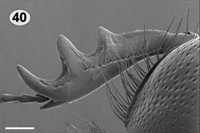
27. Protibia with teeth equally separated (Fig. 40); Argentina ................................................................ Orodaliscoides reflexus (Schmidt)
- Protibia with apical 2 teeth (1+2) closer than next pair (2+3) (Fig. 41); Chile
.............................................................. Orodaliscoides rugosiceps (Harold)

Fig. 40. Orodaliscoides reflexus protibia; Fig. 41. Orodaliscoides
rugosiceps
protibia. Scale line = 0.2 mm.
.

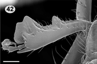
28. Protibial apex modified, bluntly expanded, apical teeth not large (Fig. 42); body dark tan to light brown, abdomen not orange .... Symphodon anomalus (Harold)

- Protibial apex not modified, not bluntly expanded, apical teeth normally developed (Figs. 63–64); body black with orange abdomen
........... Podotenus (Pseudopodotenus) fulviventris (Fairmaire and Germain)

Fig. 42. Symphodon anomalus protibia; Fig. 63. Podotenus fulviventris female protibia;
Fig. 64. Podotenus fulviventris male protibia. Scale line = 0.2 mm.
.
Go to start of key
.

Authors: Andrew Smith ( Canadian Museum of Nature) and
Paul Skelley (Florida Department of Agriculture and Consumer Services).
This website is based upon work supported by the National Science Foundation under Grant No.0342189.
Generated on: 28/NOV/07.....Last modified: 28/NOV/07

site designed by Aura Paucar-Cabrera