| 1. . |
Pronotum with basal bead absent, only lightly impressed near the posterior angles | |
| 1'. |
Pronotum with basal bead present, complete or nearly complete (occasionally interrupted at middle) | |
| 2(1). . . . |
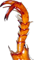
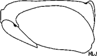
Sternites 2-4 with posterior border deeply sinuate (Fig. 11). Pygidial disc of male (in lateral view) strongly, acutely protuberant (Fig. 12). Elytral epipleuron of female weakly arcuate at mexacoxae (ventral view) (Fig. 13) | |||||||||||||||||||
| 2'. |
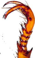
Sternites 2-4 with posterior border straight or weakly sinuate (Fig. 14). Pygidial disc of male (in lateral view) not protuberant (Fig.15). Elytral epipleuron of female straight or notched, not arcuate at metacoxa (ventral view) (Fig. 16) | |||||||||||||||||||
|
||||||||||||||||||||
| 3(2). . |
Mesosternal process with apex rounded. Sternum with dense setae. Dorsal color yellow-brown or yellow-orange | . 4 |
| 3'. |
Mesosternal process with apex acute. Sternum with scarce setae. Dorsal color dark red or mahogany with margins of the pronotum and elytral yellow | |
| 4(3). . . |
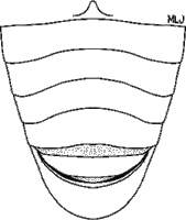
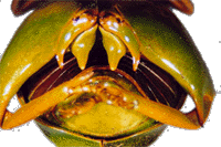
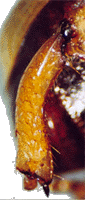
Mentum with anterior border sinuate (Fig.17). Sexual dimorphism not distinct. Posterior border of metafemur (male) without medial spine | |||||
| 4'. |
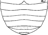
Mentum with anterior border emarginate (Fig. 18). Sexual dimorphism distinct. Posterior border of metafemur (male) with medial spine | |||||
|
||||||
| 5(4). . . . |
Metatibia of male with dense setae on the inner margin (Fig. 19). Parameres fused at mid-base. Females with elytral epipleuron parallel throughout, not convergent at apex (ventral view) (Fig.20). Mountains of Chiapas (Mexico) and Guatemala | |||||||||||||
| 5'. |
Metatibia of male with scarce setae on the inner margin (Fig. 21). Parameres not fused at mid-base. Females with elytral epipleuron convergent at apex (ventral view) (Fig. 22). Mountains of Durango, Nayarit, and Jalisco (Mexico) | |||||||||||||
|
||||||||||||||
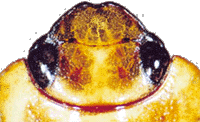
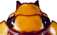
| 6(1). . |
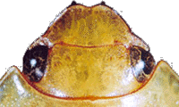
Anterior border of clypeus sinuate (Fig. 22, Fig. 23, Fig. 24, Fig. 25) | ||||||||||||||
| 6'. |
Anterior border of clypeus rounded (Fig. 26) | ||||||||||||||
|
|||||||||||||||
| 7(6). . |
Dorsal color green or olive-green without longitudinal markings. Metacoxae of male with long, apical spine (Fig. 27) | |
| |
||
| 7'. |
Dorsal color tan, cream, or yellowish with or without longitudinal markings. Metacoxae of male without apical spine | |
| 8(7). . |
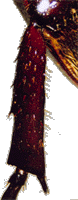
Metatibia of male with long, inner spine. Body length 30-35 mm | |
| 8'. |
Metatibia of male without inner spine. Body length 15-20 mm | |
| 9(8). . |
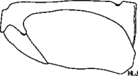
Metatrochanter with apex not produced beyond posterior border of femur (Fig. 28). Metatibia of male straight (Fig. 29) | . 10 |
||||||||||||
| 9'. |
Metatrochanter with apex weakly spine-like and produced beyond posterior border of femur (Fig. 30). Metatibia of male curved (Fig. 31) | |||||||||||||
|
||||||||||||||
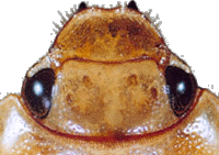
| 10(9). . . |
Clypeus near apex punctostriate (Fig. 23). Mesotarsomeres of male thickened and foreshortened (Fig. 32). Modified mesostarsal claw of male wider than modified protarsal claw; apex bulbous and widely split (Fig. 32) | |||||||||||||
| 10'. |
Clypeus near apex punctate or rugopunctate (Fig. 24). Mesotarsomeres of male not thickened and foreshortened (Fig. 33). Modified mesotarsal claw of male subequal in width to modified protarsal claw; apex not bulbous and widely split (Fig. 33) | |||||||||||||
|
||||||||||||||