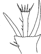
| 1. |
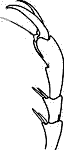
Antennal club 3-8 segmented, symmetrical, usually lamellate (figs. 1-7). Head not covered by prothorax. Forecoxae large, strongly transverse or conical and projecting below prosternum. Foretibiae flattened with one or more teeth on outer edge. Tarsi with 5 distinct segments, none of which is lobed or densely pubescent | |||||||||||||||
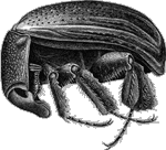
Figures
1-7. Right antenna dorsal view of: (1) Diphyllostoma
sp. (Diphyllostomatidae), (2) Platycerus
sp. (Lucanidae), (3) Odontotaenius disjunctus
(Passalidae), (4) Hybosorus illigeri (Hybosoridae),
(5) Pleocoma sp. (Pleocomidae), (6)
Bradycinetulus sp. (Geotrupidae), (7)
Euphoria sp. (Scarabaeidae). |
||||||||||||||||


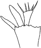
| 2 (1). | Antennae with 11 segments (figs. 5,6) | |||||||||||
Figures
5-6. Right antenna dorsal view of: (5) Pleocoma
sp. (Pleocomidae), (6) Bradycinetulus
sp. (Geotrupidae). |
||||||||||||
| 2'. | Antennae with fewer than 11 segments (figs. 1-4, 7) | |||||||||||
|
||||||||||||
| 3(2). | Antennal club with 4-7 elongate segments (fig. 5) | Pleocomidae | |
 Figure 5. Right antenna dorsal view of: Pleocoma sp. (Pleocomidae). |
|||
| 3'. | Antennal club with 3 circular or oval segments (fig. 6) | Geotrupidae | |
Figure 6. Right antenna dorsal view of: Bradycinetulus sp. (Geotrupidae). |
|||
| 4(2). . |
Body capable of being rolled into a contracted sphere (fig. 8). Middle and posterior tibiae flattened and dilated | Hybosoridae- Ceratocanthinae |
Figure
8. Ceratocanthus sp. lateral view (Ceratocanthidae). |
||
| 4'. . |
Body oblong, not capable of being rolled into a sphere. Middle and posterior tibiae not significantly flattened and dilated | |
| 5
(4). |
Mesotibia at apex with longer spur pectinate along one edge (fig. 9) | Ochodaeidae |
Figure 9. Spur at apex of mesotibia of Ochodaeus sp. (Ocodaeidae) (pectinate). |
||
| 5'. | Mesotibia at apex with spurs simple, not pectinate (fig. 10) | |
| Fig. 10. Spur at apex of mesotibia of Pleocoma sp. (Pleocomidae) (simple). |
||
| 6
(5). . |
Segments of antennal club not capable of being tightly closed together (figs. 1-3) | |||||||||
|
||||||||||
| 6' | Segments of antennal club capable of being closed together (figs. 4-7) | |||||||||
|
||||||||||
| 7
(6). . |
Mentum deeply emarginate (fig. 11). Head often with central, anterior horn (fig. 12) | |||||
|
||||||
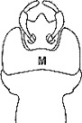
| 7'. |
Mentum simple, not deeply emarginate (fig. 13). Head without central horn | |||||

Figure 13. Ventral view of mentum (M) of Lucanus sp. (Lucanidae) (apex simple, truncate). |
||||||
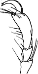
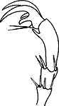
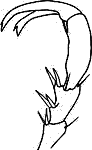
| 8
(7). . |
First antennal segment much longer than segments 2 and 3 together (figs. 2, 14). Antenna geniculate (fig. 2) (exception: straight or weakly geniculate in Ceruchus, fig. 14) | |||||
|
||||||
| 8'.
. |
First antennal segment subequal to segments 2 and 3 together (fig. 1). Antenna not geniculate (fig. 2) | Diphyllostomatidae |
||||
|
||||||
| 9
(6). . |
Antennal club with 3 segments, first segment hollowed out to receive second segment (fig. 4) | Hybosoridae KEY |
 Figure 4. Right antenna dorsal view of Hybosorus illigeri (Hybosoridae). |
||
| 9'.
. |
Antennal club with 3-7 segments, first segment simple, not hollowed out to receive second segment (e.g., fig. 7) | |

Figure 7. Right antenna dorsal view of Euphoria sp. (Scarabaeidae). |
||
| 10
(9). . |
Abdomen with 5 ventral sclerites (fig. 15). Dorsal surface roughened or tuberculate, not shining | |
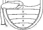 Figure 15. Abdomen and posterior leg of Omorgus sp. (Trogidae). |
||
| 10'.
. |
Abdomen with 6 ventral sclerites (fig. 16). Dorsal surface variably sculptured, shining or not | |
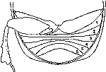 Figure 16. Abdomen and posterior leg of Copris sp. (Scarabaeidae). |
||
| 11
(10). |
Antenna 9-segmented | |
| 11'.
|
Antenna 10-segmented | |
| |
||
| 12
(11). . . . |
Eyes not divided by canthus (fig. 17). Clypeus with sides narrowing to apex. Color brown, gray, or black. Metafemora and metatibia not enlarged, not covering abdomen | |
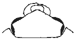 Figure 17. Dorsal view of head of Omorgus (eyes not divided by canthus). |
||
| 12'. . . . |
Eyes divided by prominent canthus (fig. 18). Clypeus with sides subparallel to divergent before apex. Color testaceous to light reddish brown. Metafemora and metatibia enlarged, covering most of abdomen | |
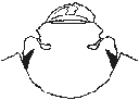 Figure 18. Dorsal view of head of Glaresis (eyes divided by prominent canthus). |
||
| 13
(10). . . |
Elytra shortened and widely divergent at apex, not covering pygidium (fig. 19) (except in L. lupina). Eighth abdominal segment with spiracle | |
 Figure 19. Lichnanthe rathvoni LeConte (Glaphyridae). |
||
| 13'.
. |
Elytra not shortened and widely divergent at apex, pygidium exposed or not. Eighth abdominal segment lacking spiracle | |
| 14
(13). . |
Pygidium
completely (or nearly so) covered by apex of elytra. Length 1.5-13.0 mm |
|
| 14'. | Pygidium completely exposed. Length longer than 5.0 mm | |
| |
||
| 15
(14). . |
Antennal insertion visible from above (clypeus with sides constricted medially just before eyes) (fig. 20) | |

Figure 20. Head and antenna (dorsal view) of Euphoria sp. showing clypeal sides constricted and with antennal insertion visible. |
||
| 15'. . . |
Antennal insertion not visible from above (clypeus with sides not constricted) | |
| 16
(15). . |
Abdominal sternites distinctly narrowed at midline (fig. 16); length of all sternites shorter than length of metasternum. Scutellum usually hidden | |
| Figure 16. Abdomen and posterior leg of Copris sp. (Scarabaeinae). |
||
| 16'. . . |
Abdominal sternites normal, not narrowed at midline; length of all sternites longer than length of metasternum. Scutellum usually visible | |
| 17
(16). . . |
Claws of both middle and posterior tarsi unequal in length and independently movable (fig. 25) (exception: all legs in Leptohoplia with only claw or with one claw greatly reduced) | |||||||||
| 17'. . . |
Claws of both middle and posterior tarsi equal in length and not independently movable (figs. 26-28) (exception: posterior tarsi in Hoplia with only one claw) | |||||||||
Figures 25-28. (25) Claws of posterior tarsi of Anomala sp. (Rutelinae) (claws simple and unequal in length). Claws of posterior tarsi of: (26) Xyloryctes jamaicensis (Dynastinae) (claws simple and equal in length), (27) Polyphylla sp. (Melolonthinae), (28) Dichelonyx sp. (Melolonthinae) (claws cleft or toothed and equal in length). |
||||||||||
| 18
(17). . . . |
Claws of middle and posterior tarsi simple (fig. 26). Base of pronotum and elytra subequal in width. Apex of posterior tibia always with 2 spurs. Mandibles often exposed in dorsal view | |||||||
| 18'. . . . . |
Claws of middle and posterior tarsi cleft, toothed (figs. 27-28), or simple (if simple, base of pronotum much narrower than base of elytra). Apex of posterior tibia with 1-2 spurs or spurs absent. Mandibles hidden in dorsal view | Melolonthinae KEY |
||||||
Figures
26-28. Claws of posterior tarsi of: (26) Xyloryctes jamaicensis
(Dynastinae) (claws simple and equal in length), (27)
Polyphylla sp. (Melolonthinae), (28) Dichelonyx
sp. (Melolonthinae) (claws cleft or toothed and equal in length).
|
||||||||
| 19
(18). . . |
Mandibles and labrum projecting anteriorly beyond clypeus, visible in dorsal view. Metatibial spines separated by base of tarsomere 1 (fig. 29) | |||||
| 19'. . . |
Mandibles and labrum not projecting anteriorly beyond clypeus, not visible in dorsal view. Metatibial spines adjacent, not separated by base of tarsomere 1 (fig. 30) | |||||
|
||||||
| 20
(19). . . |
Prosternal projection (posterior to procoxae) produced to level of procoxae, conical. Venter with abundant, long, tawny setae | |
| 20'. . . |
Prosternal projection (posterior to procoxae) triangular, produced to middle of procoxae. Venter without abundant, long setae | |