Males: Protarsomeres dorsoventrally flattened,
densely setose ventrally (Figs. 30-31, 33-34); terminal sternite with
margin emarginated; abdominal sternites in lateral view appearing concave
or flat. Females: Protarsomeres dorsoventrally flattened
or not, with or without dense ventral setae; terminal sternite with margin
entire or rounded, not emarginated; abdominal sternites in lateral view
appearing convex. |
1.
|
Mentum with
apicomedial, tooth-like projection (Fig. 16, 41) |
|
|
 Trizogeniates
foveicollis
Trizogeniates
foveicollis |
Figure
16 |
Figure
41 |
Figure
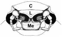
16. Geniatini and Anoplognathini with labrum and mentum (each)
possessing median, apical tooth or projection. Figure 41.
Trizogeniates foveicollis (clypeal apex with apicomedial
tooth-like projection). |
| 1'.
|
Mentum
without apicomedial, tooth-like projection (Figs. 38-40) |
|
Mimogeniates margaridae |
Rhizogeniates antennatus |
Rhizogeniates
carbonarius
|
Figure
38 |
Figure
39 |
Figure
40 |
Figures
38-40. Clypeus of: 38)Mimogeniates
margaridae (apex quadrate and crenulate, lacking apicomedial
tooth), 39) Rhizogeniates antennatus (weakly
emarginate apically, lacking apicomedial tooth), 40)
Rhizogeniates carbonarius (weakly emarginate apically,
lacking apicomedial tooth. |
|
| |
| |
| |
| |
| |
| |
| |
| |
| |
| |
| |
| |
| |
| |
| |
| |
| |
| |
| |
| |
| |
| |
| |
| |
| |
|
2(1).
. |
Apex of mentum
with medial notch, not crenulate (Figs. 39-40). All claws simple on
all legs |
|
| |
| 2'.
. |
Apex
of mentum crenulate (Fig. 38). Modified claw moderately split on all
legs |
|
Mimogeniates
margaridae
|
Rhizogeniates
antennatus
|
Rhizogeniates
carbonarius
|
|
Figure 38 |
Figure
39 |
Figure
40 |
Figures
38-40. Clypeus of: 38)Mimogeniates
margaridae (apex quadrate and crenulate, lacking apicomedial
tooth), 39) Rhizogeniates antennatus (weakly
emarginate apically, lacking apicomedial tooth), 40)
Rhizogeniates carbonarius (weakly emarginate apically,
lacking apicomedial tooth. |
|
| |
| |
| |
| |
| |
| |
| |
| |
| |
| |
| |
| |
| |
| |
| |
| |
| |
| |
| |
| |
| |
| |
| |
| |
| |
| |
| |
|
3(1).
. |
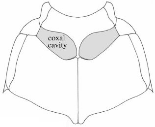
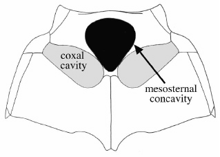
Mesosternum
anterior to mesocoxae strongly concave (Fig. 52) |
|
| |
| 3'.
. |
Mesosternum
anterior to mesocoxae flat or slightly convex, not strongly concave
(Fig. 51) |
|
 Bolax
magna
Bolax
magna |
 Xenogeniates
martinezi
Xenogeniates
martinezi |
Figure
51 |
Figure
52 |
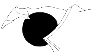
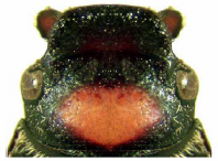
Figure
51-52.
Thorax in ventral view showing: 51) Mesosternum
without invagination (Bolax magna) and 52) Mesosternum
with invagination in black (Xenogeniates martinezi). |
|
| |
| |
| |
| |
| |
| |
| |
| |
| |
| |
| |
| |
| |
| |
| |
| |
| |
| |
| |
| |
| |
| |
| |
| |
| |
| |
| |
|
4(3).
. |
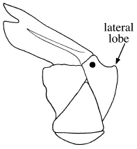
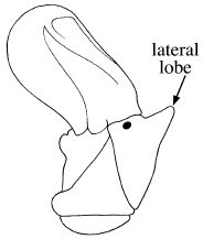
Stipes of maxilla
produced, with well-developed lateral lobe (Fig. 43) or lateral angle
(Fig. 42)
|
|
| |
| 4'.
. |
Stipes
of maxilla not produced, instead rounded or broadly rounded (Fig.
44) |
|
 Lobogeniates
borgmeieri Lobogeniates
borgmeieri |
 Lobogeniates
catullus Lobogeniates
catullus |
 Trizogeniates
foveicollis
Trizogeniates
foveicollis |
Figure
42 |
Figure
43 |
Figure
44 |
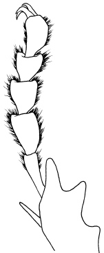
Figure
42-44. Maxilla (ventral view) showing: 42) Lobogeniates
borgmeieri (stipes produced, with lateral angle), 43)
Lobogeniates catullus (stipes produced, with well-developed lateral
lobe), 44) Trizogeniates foveicollis (not
produced, instead rounded or broadly rounded). |
|
| |
| |
| |
| |
| |
| |
| |
| |
| |
| |
| |
| |
| |
| |
| |
| |
| |
| |
| |
| |
| |
| |
| |
| |
| |
| |
| |
| |
| |
| |
|
5(4).
.
.
. |
Mandible with
rounded, recurved, apical lobe (Fig. 20). Dorsal surface with abundant,
decumbent, white setae. Antennal club of male twice length of segments
2-7; antennal club of female subequal to segments 2-7 |
|
| |
| 5'.
.
.
. |
Mandible
lacking rounded, recurved, apical tooth; instead simple (e.g., Fig.
19). Dorsal surface with or without sparse setae. Antennal club of
male and female subequal to or slightly longer than segments 2-7 |
|
 Bolax
rutila
Bolax
rutila |
 Eunanus
murinus
Eunanus
murinus |
Figure
19 |
Figure
20 |
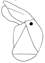
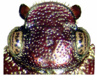
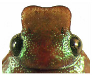
Figures
19-20.
Head in dorsal view showing form of clypeus, mouthparts, and eye size
in: 19) Bolax rutila, 20)
Eunanus murinus. |
|
| |
| |
| |
| |
| |
| |
| |
| |
| |
| |
| |
| |
| |
| |
| |
| |
| |
| |
| |
| |
| |
| |
| |
| |
| |
| |
| |
|
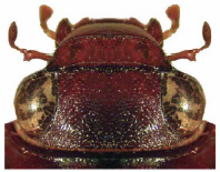
6(5).
.
.
. |
Length of antennal
club half or less than half length of first antennal segment (Figs.
5, 23). Clypeal apex (in lateral view) sloped 45º with respect to
dorsal plane of clypeus (Figs. 23, 36). Male tarsomeres simple, not
flattened and dilated (Fig. 32) |
|
| |
| 6'.
.
.
. |
Length
of antennal club more than half length of first antennal segment.
Clypeal apex (in lateral view) sloped 60-90º with respect to dorsal
plane of clypeus (Fig. 37). Male tarsomeres dorsoventrally flattened
and dilated (e.g., Fig. 34) |
|
|
|
|
|
Figure
23 |
Figure
32 |
Figure
34 |
|
|
|
Figure
5 |
Figure
36 |
Figure
37 |
|
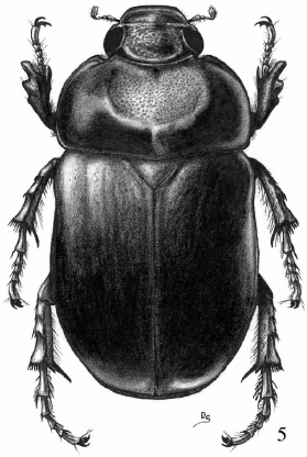
Figure 5. Geniatosoma nigrum (male).
Figure 23. Head in dorsal view showing form of
clypeus, mouthparts, and eye size in Geniatosoma lindemannae.
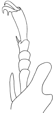
Figures 32, 34. Male forelegs (dorsal view) showing comparison
of tibial apex, protarsomeres, and claws of: 32) Geniatosoma
lindemannae, 34) Trizogeniates temporalis. Figures
36-37. Head and apex of thorax in lateral view showing:
36) Clypeal apex sloped 45º with respect to dorsal
plane of clypeus in Geniatosoma lindemannae (male) and 37)
Clypeal apex sloped 60–90º with respect to dorsal plane of
clypeus in Trizogeniates tibialis. |
|
| |
| |
| |
| |
| |
| |
| |
| |
| |
| |
| |
| |
| |
| |
| |
| |
| |
| |
|
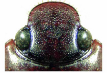
7(6).
.
.
|
Form
of clypeus parabolic, apex not reflexed (Fig. 24). Mandible
exposed, apex narrowly rounded (Fig. 24). Male with all claws
appearing simple on all legs |
|
| |
| 7'.
.
.
. |
Form
of clypeus not parabolic (instead rounded, quadrate), apex reflexed
(e.g., Figs. 19-20, 22, 25-26). Mandible exposed or not, apex
broadly rounded (e.g., Figs. 19, 23, 25). Male with claws obviously
toothed on some or all legs |
|
Bolax
rutila |
Eunanus
murinus |
 Geniates
borellii
Geniates
borellii |
 Geniatosoma
lindemannae
Geniatosoma
lindemannae |
Figure
19 |
Figure
20 |
Figure
22 |
Figure
23 |
 Heterogeniates
bonariensis Heterogeniates
bonariensis
|
 Leucothyreus
kirbyanus Leucothyreus
kirbyanus
|
 Trizogeniates
tibialis Trizogeniates
tibialis
|
|
| Figure
24 |
Figure
25 |
Figure
26 |
|
Figures
19-26. Head in dorsal view showing form of clypeus,
mouthparts, and eye size in: 19) Bolax
rutila, 20) Eunanus murinus,
22) Geniates borellii, 23)
Geniatosoma lindemannae, 24)
Heterogeniates bonariensis (male), 25)
Leucothyreus kirbyanus, 26) Trizogeniates
tibialis. |
|
| |
| |
| |
| |
| |
| |
| |
| |
| |
| |
| |
| |
| |
| |
| |
| |
| |
| |
| |
| |
| |
| |
| |
| |
| |
| |
| |
|
| 8(7).
.
.
.
|
Length
of protarsomeres 2-4 subequal in length to protarsomere 5 (Fig.
31). Clypeus of male with lateral margins expanded, apex quadrate
(Fig. 21); clypeus of female with lateral margins parallel,
apex quadrate |
|
| |
| 8'.
.
.
. |
Length
of protarsomeres 2-4 greater than length of protarsomere 5 (Figs.
30, 33-34). Clypeus of male and female with lateral margins
constricted, apex rounded or trapezoidal (e.g., Figs. 19, 22,
25-28) |
|
Bolax
|
 Evanos Evanos
|
Geniates
|
Leucothyreus
|
Figure
19 |
Figure
21 |
Figure
22 |
Figure
25 |
Trizogeniates
|
 Geniates
borellii Geniates
borellii
|
 Geniates
cornutus Geniates
cornutus
|
|
Figure
26 |
Figure
27 |
Figure
28 |
|
Bolax
|
Evanos
|
Microchilus
|
Trizogeniates
|
Figure
30 |
Figure
31 |
Figure
33 |
Figure
34 |
Figures
19-26. Head in dorsal view showing form of clypeus,
mouthparts, and eye size in: 19) Bolax
rutila, 21) Evanos villatus (male),22)
Geniates borellii, 25)
Leucothyreus kirbyanus, 26) Trizogeniates
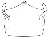
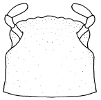
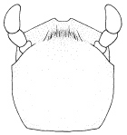
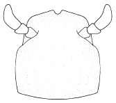
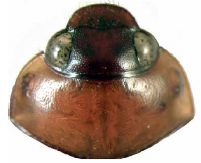
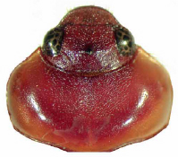
tibialis. Figures 27-28. Head and
pronotum in dorsal view showing form in: 27) Geniates
borellii, male (head lacking tubercle, pronotum lacking
concavity), 28) Geniates cornutus,
male (head with tubercle, pronotum with concavity).
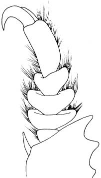
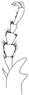
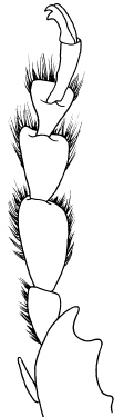
Figures 30-34. Male forelegs (dorsal view)
showing comparison of tibial apex, rotarsomeres, and claws
of: 30) Bolax magna, 31) Evanos villatus,
33) Microchilus lineatus, and 34) Trizogeniates
temporalis.
|
|
| |
| |
| |
| |
| |
| |
| |
| |
| |
| |
| |
| |
| |
| |
| |
| |
| |
| |
| |
| |
| |
| |
| |
| |
| |
| |
| |
| |
| |
|
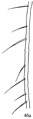
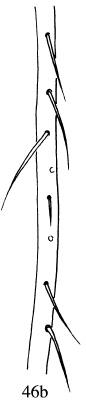
9(8).
. |
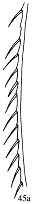
Elytral
margin with deep, setose punctures on lateral edge from apex
of metepisternum to apex of elytra (Figs. 45a-b, 46a-b) |
|
| |
| 9'.
. |
Elytral
margin without deep, setose punctures on lateral edge from apex
of metepisternum to apex of elytra |
|
|
|
|
|
Trizogeniates
foveicollis |
Geniates
cylindricus |
Figure
45 |
Figure
46 |
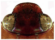
Figures
45-46. Left elytral epipleuron showing: 45)
dorsal view of stridulatory ridge in T. foveicollis
(a) and ventral view of stridulatory ridge
in T. foveicollis (b), 46)
dorsal view of elytral epipleuron without stridulatory ridge
in G. cylindricus (a) and ventral
view of elytral epipleuron without stridulatory ridge in G.
cylindricus (b). |
|
| |
| |
| |
| |
| |
| |
| |
| |
| |
| |
| |
| |
| |
| |
| |
| |
| |
| |
| |
| |
| |
| |
| |
| |
| |
| |
| |
| |
| |
|
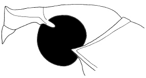
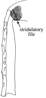
10(9).
.
. |
Elytral
margin with well-developed stridulatory ridge and with rigid
stridulatory setae (Fig. 45a-b). Apex of metafemur (dorsal view)
with stridulatory patch (Fig. 35) |
|
| |
| 10'.
.
. |
Elytral
margin lacking stridulatory ridge and without rigid stridulatory
setae (Fig. 46a-b). Apex of metafemur (dorsal view) lacking
stridulatory patch |
|
|
 |
|
|
|
Figure
35 |
Figure
45 |
Figure
46 |
Figure
35. Hind leg (dorsal view) of Trizogeniates temporalis
showing location of stridulatory file at the apex of the metafemur.Figures
45-46. Left elytral epipleuron showing: 45)
dorsal view of stridulatory ridge in T. foveicollis
(a) and ventral view of stridulatory ridge
in T. foveicollis (b), 46)
dorsal view of elytral epipleuron without stridulatory ridge
in G. cylindricus (a) and ventral
view of elytral epipleuron without stridulatory ridge in G.
cylindricus (b). |
|
| |
| |
| |
| |
| |
| |
| |
| |
| |
| |
| |
| |
| |
| |
| |
| |
| |
| |
| |
| |
| |
| |
| |
| |
| |
| |
| |
| |
| |
| |
| |
| |
|
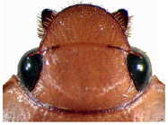
11(9).
. |
Eyes
small, interocular width greater than 6 transverse eye diameters
(e.g., Fig. 19) |
|
| |
| 11'.
. |
Eyes
larger, interocular width less than 5 transverse eye diameters
(e.g., Fig. 25) |
|
Bolax
|
Leucothyreus
|
Figure
19 |
Figure
25 |
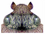
Figures
19, 25. Head in dorsal view showing form of clypeus,
mouthparts, and eye size in: 19) Bolax
rutila, 25) Leucothyreus kirbyanus,
|
|
| |
| |
| |
| |
| |
| |
| |
| |
| |
| |
| |
| |
| |
| |
| |
| |
| |
| |
| |
| |
| |
| |
| |
| |
| |
| |
| |
| |
| |
|
12(11).
.
. |
Protarsomere
5 dorsoventrally flattened, width more than half length (Fig.
30). Length of body from apex of clypeus to apex of elytra more
than 9.0 mm |
Bolax
Fischer von Waldheim
|
Figure
30. Male
foreleg (dorsal view) showing comparison of tibial apex, rotarsomeres,
and claws of Bolax magna |
| 12'.
.
. |
Protarsomere
5 dorsoventrally flattened or not; if flattened, then width
less than half length. Length of body from apex of clypeus to
apex of elytra less than 9.0 mm |
|
|
| |
| |
| |
| |
| |
| |
| |
| |
| |
| |
| |
| |
| |
| |
| |
| |
| |
| |
| |
| |
| |
| |
| |
| |
|