| 1. . |
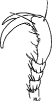
Labrum horizontally produced with respect to clypeal apex (Fig. 1), distinctly separated from clypeus | . 2 |
||||||
| 1'. |
Labrum vertically produced with respect to clypeal apex (Fig.2) and more or less fused to clypeus | |||||||
|
||||||||

| 2(1). . |
Margin of elytra with membranous border. Antenna 9-segmented | |
| 2'. |
Margin of elytra without membranous border. Antenna 10-segmented (except in Parachrysina (Areodina), Eremophagous (Pelidnotina), and female Pseudogeniates richterianus (Pelidnotina)] | |
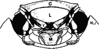
| 3(1). . |
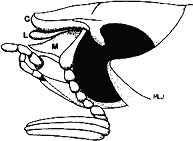
Mentum
and labrum each with median, apical tooth or projection (Fig. 3) |
. 5 |
||||||||||
| 3'. . |
Mentum lacking median, apical tooth or projection; labrum with or without median, apical tooth or projection (Figs. 4, 5) | . 4 |
||||||||||
C=clypeus, L=labrum, M=mentum |
||||||||||||
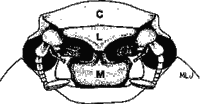
| 4(3). . |
Labrum with median, apical projection; apex overhanging mentum (Fig.4) | . Adoretini |
||||||
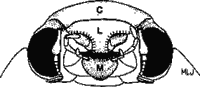
| 4'.
|
Labrum and mentum both simple, lacking median, apical projection (Fig. 5) | . Spodochlamyini |
||||||
C=clypeus, L=labrum, M=mentum |
||||||||
| 5
(3). . |
Protarsomeres dorsoventrally flattened and expanded apically in males and/or males and females (Fig. 6) | |||||||
| 5'. |
Protarsomeres simple, not dorsoventrally flattened and expanded apically in males or females (Fig. 7) | |||||||
|
||||||||